Transcriptomic & Immune Microenvironment Profiling of Bladder Cancer Progression
A precision-medicine pipeline in differential expression, pathway enrichment, and immune deconvolution across 233 bladder tissue samples.
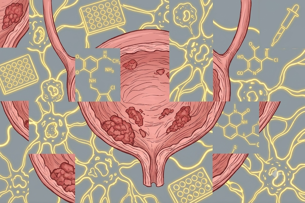
At a glance
The biology separating a contained bladder tumor from one that invades is unresolved, yet that single distinction drives entirely different treatment paths.
A four-stage R pipeline — differential expression, functional enrichment, immune deconvolution, and longitudinal visualization — tracing molecular and immune change from pre-cancer to muscle-invasive disease.
Surfaced an EMT/ECM remodeling signature at tumor onset and a TNF-driven inflammatory shift at invasion, plus immune-composition changes relevant to immunotherapy timing and patient selection.
Overview
This project traces the molecular and immune changes that accompany bladder cancer as it moves from pre-cancerous tissue through non-muscle-invasive (stage 1) to muscle-invasive (stage 2) disease. Bladder cancer is staged as non-muscle-invasive or muscle-invasive, and that single distinction drives very different treatment paths — intravesical therapy versus systemic chemotherapy or cystectomy. Roughly half of non-muscle-invasive tumors recur, yet the biology separating a contained tumor from an invasive one is not fully resolved.
Using a public Illumina microarray cohort (GSE13507, Kim et al.) of 233 bladder tissue samples — 10 normal mucosa, 58 surrounding pre-cancerous field, 103 superficial stage 1, and 62 invasive stage 2 — with matched clinical annotation for stage, recurrence, and progression, I built a four-stage analytical pipeline in R that identifies differentially expressed genes between disease stages, maps them to biological pathways, estimates the immune-cell composition of each tumor, and tracks how that composition shifts across the progression trajectory.
Phase 1 — Differential expression: a custom fnTTest function runs a Welch two-sample t-test per probe with signed log2 fold change and Benjamini-Hochberg FDR correction. The pre-cancer→stage 1 transition produced the strongest signal, with stromal/ECM genes (MFAP4, CFD, COL16A1, DCN, ACTG2) sharply downregulated (FDR ~1e-20 or smaller); the stage 1→stage 2 comparison was more modest, led by UNC5B, KRTAP5-2, and CRTAC1.
Phase 2 — Functional enrichment: the top 1,000 DEGs per comparison were submitted to EnrichR across ~30 curated libraries (KEGG, GO BP, Reactome, WikiPathways, BioPlanet, MSigDB Hallmark), compiled into per-comparison Excel workbooks. EMT was the single most enriched Hallmark pathway in both transitions, alongside TGF-beta ECM regulation and matrix organization. TNF-alpha signaling via NF-kB became prominent specifically at the transition to invasion — a distinct inflammatory program layered on ongoing matrix remodeling.
Phase 3 — Immune deconvolution: CIBERSORTx against the LM22 signature (22 immune subsets, quantile normalization disabled per microarray guidance), run per tissue group and stratified by recurrence. Normal mucosa was dominated by naive/memory B cells (24%/16%) and CD8 T cells (11%), while tumor tissue was dominated by neutrophils (54%) and eosinophils (16%) — a shift from adaptive surveillance toward innate, myeloid-driven infiltration. Across the trajectory, Tregs rose from ~7.5% (pre-cancer) to 13.5% (stage 1) then fell to ~9% (stage 2), activated dendritic cells rose from ~5% to ~13% and stayed elevated, and M2 macrophages declined steadily from ~12% to ~9%.
Phase 4 — Longitudinal visualization: a reusable functionLineGraph reshapes the 22 cell-type fractions and plots each immune subset across the progression axis and recurrence status with ggplot2, exporting publication-ready PNG, TIFF, and PDF figures. Together the evidence points to an immunosuppressive shift at the earliest steps of transformation and a more immune-quiescent microenvironment in fully invasive disease — a compact demonstration of pushing a single public dataset through a genuine precision-medicine workflow, from raw expression values to a testable hypothesis about immune evasion.
Highlights
- Genome-scale differential expression: per-probe Welch t-tests across ~43,000 probes with Benjamini-Hochberg FDR correction
- EMT and ECM remodeling identified as the dominant signature at tumor onset (MSigDB Hallmark, BioPlanet, Reactome)
- TNF-alpha / NF-kB inflammatory signaling flagged specifically at the transition to muscle-invasive disease
- CIBERSORTx immune deconvolution revealing rising Tregs and dendritic cells early, then a more immune-quiescent invasive microenvironment